Home
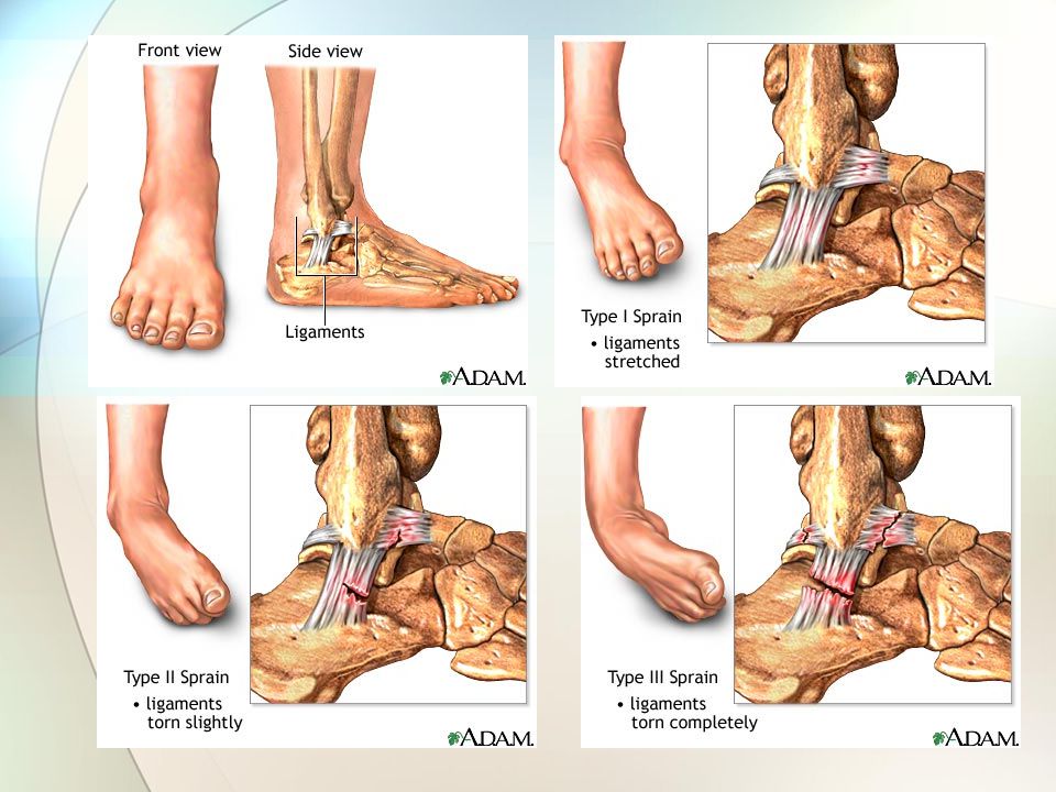
/ Inferior Extensor Retinaculum Swelling - What Is Tendonitis, Retinaculum tibia tendon of tibialis anterior muscle medial malleolus inferior extensor retinaculum tendon of extensor hallucis longus muscle (sheath).
Inferior Extensor Retinaculum Swelling - What Is Tendonitis, Retinaculum tibia tendon of tibialis anterior muscle medial malleolus inferior extensor retinaculum tendon of extensor hallucis longus muscle (sheath).
Inferior Extensor Retinaculum Swelling - What Is Tendonitis, Retinaculum tibia tendon of tibialis anterior muscle medial malleolus inferior extensor retinaculum tendon of extensor hallucis longus muscle (sheath).. At 30º plantar flexion, the talonavicular joint was hypermobile and the extensor digitorum longus tendon was impinged on by the talar head and slipped laterally. The inferior extensor retinaculum (ier) is an aponeurotic structure, which is in continuation with the anterior part of the sural fascia. The dpn is most commonly entrapped at the inferior edge of the extensor retinaculum where the extensor hallucis brevis crosses over top. 3) weakness of extensor hallucis longus and extensor digitorum communis; 2) hypoaesthesia or anaesthesia in the web space of the great toe;
And 4) pain on passive flexion of the toes, especially the great toe. Person suffering from inferior extensor retinaculum pain or strain may have symptoms of difficulty in wearing shoes and socks with experience of pain in the foot and ankle. Retinaculum tibia tendon of tibialis anterior muscle medial malleolus inferior extensor retinaculum tendon of extensor hallucis longus muscle (sheath). Symptoms are likely to develop gradually over time, becoming progressively worse. Weakness and atrophy of the edb may occur if compression occurs at the superior edge.
Extensor Tendonitis Treatment In North Texas Graff Foot Ankle And Wound Care from drgraff.com You may have some diffuse swelling over the top of your foot. Symptoms are likely to develop gradually over time, becoming progressively worse. There is a physiological oedema occurring during sleep in every human cornea amounting to an increase in thickness of about 4%. Peroneal retinacula are often injured during ankle sprains (inversion injury). It is directed medialward as a double layer, one lamina passing in front of, and the other behind, the tendons of the peroneus tertius and extensor digitorum longus. Weakness and atrophy of the edb may occur if compression occurs at the superior edge. Moderate to severe pain in the ankle and foot with ambulating or running or in fact any movement of the foot. A study published in american journal of roentgenology in 2006 studied the superior extensor retinaculum and surrounding tissues of cadavers, and found that injuries to the superior extensor.
3) weakness of extensor hallucis longus and extensor digitorum communis;
There are several potential sites of entrapment, which result in slightly different clinical presentations. It is directed medialward as a double layer, one lamina passing in front of, and the other behind, the tendons of the peroneus tertius and extensor digitorum longus. Anytime there is persistent pain after an ankle sprain, a retinaculum injury should be considered. We describe six patients aged from 10 to 15 years who, after injury to the distal tibial physis, presented with the following clinical findings: Swelling over the dorsal ankle, particularly along the extensor digitorum longus tendon. 9 moreover, the inferior extensor retinaculum supplies the retinacular roots of the sinus tarsi. The pain is located at the dorsomedial aspect of the foot and is worst at rest. Originates from the calcaneus, the interosseous talocalcaneal ligament, and the inferior extensor retinaculum and inserts to the base of the proximal phalanx of the great toe. In addition there is a superior.flexor digitorum longus pain running. The anterior tarsal tunnel syndrome denotes the entrapment of the deep peroneal nerve under the inferior extensor retinaculum 3). Extensor tendons connect muscle to bone in the hand and foot, and extensor tendonitis is commonly caused by overuse. The stem, particularly the medial root, of the inferior retinaculum forms a constraining loop around the edl and peroneus tertius tendons. The superior extensor retinaculum also interacts with tendons in your lower body, so injury and pain to this ligament might have other effects.
The most common cause is overuse of the muscles, bones, and tendons in the feet. Moderate to severe pain in the ankle and foot with ambulating or running or in fact any movement of the foot. 9 moreover, the inferior extensor retinaculum supplies the retinacular roots of the sinus tarsi. There are several potential sites of entrapment, which result in slightly different clinical presentations. The anterior tarsal tunnel is a fibroosseous canal between the inferior extensor retinaculum and the talus and navicular.
Extensor Tendonitis Causes Symptoms Types 10 Faqs Answered from www.ugetwellsoon.com Moderate to severe pain in the ankle and foot with ambulating or running or in fact any movement of the foot. The dpn is most commonly entrapped at the inferior edge of the extensor retinaculum where the extensor hallucis brevis crosses over top. Originates from the calcaneus, the interosseous talocalcaneal ligament, and the inferior extensor retinaculum and inserts to the base of the proximal phalanx of the great toe. It is often described as an aching pain, which increases with exercise, in particular, running. This syndrome is secondary to dpn compression at the inferior extensor retinaculum where the extensor hallucis longus tendon crosses over it (, 22). Failure of the repair of the inferior extensor retinaculum can result in bowstringing of the tibialis anterior tendon during ankle dorsiflexion. The pain is located at the dorsomedial aspect of the foot and is worst at rest. The main symptoms indicating inferior extensor retinaculum strain are:
The extensor retinaculum is a thick band of fascia that holds the organs of the feet in their place.
The extensor retinaculum in the foot is a thick band that runs from the front of the ankle joint to the back of the foot. Other findings included a bulbous appearance or swelling of the torn tendon in two complete and two partial tears and fluid collections within the tendon sheath and in an area confined by the extensor retinaculum in four patients. Symptoms are likely to develop gradually over time, becoming progressively worse. 2,3 it results in a predominately sensory neuropathy. Weakness and atrophy of the edb may occur if compression occurs at the superior edge. Pain and swelling posterior to the. 3) weakness of extensor hallucis longus and extensor digitorum communis; The pain is located at the dorsomedial aspect of the foot and is worst at rest. Extensor hallucis longus in extending the great toe at the metatarsophalangeal joint. Inferior extensor retinaculum is a term used to describe pain in the lower extremities (feet) arising due to irritation of the retinaculum of the foot. The main symptom of extensor tendonitis is pain on the top of the foot. There is a physiological oedema occurring during sleep in every human cornea amounting to an increase in thickness of about 4%. The inferior extensor retinaculum has a more complex morphology, resembling a sideways letter y over the anterior tibotalar joint and dorsal midfoot.
9 moreover, the inferior extensor retinaculum supplies the retinacular roots of the sinus tarsi. The extensor retinaculum in the foot is a thick band that runs from the front of the ankle joint to the back of the foot. Other findings included a bulbous appearance or swelling of the torn tendon in two complete and two partial tears and fluid collections within the tendon sheath and in an area confined by the extensor retinaculum in four patients. The most common cause is overuse of the muscles, bones, and tendons in the feet. Originates from the calcaneus, the interosseous talocalcaneal ligament, and the inferior extensor retinaculum and inserts to the base of the proximal phalanx of the great toe.
The Ankle And Foot Joints Ppt Download from images.slideplayer.com Moderate to severe pain in the ankle and foot with ambulating or running or in fact any movement of the foot. Symptoms are likely to develop gradually over time, becoming progressively worse. The extensor retinaculum in the foot is a thick band that runs from the front of the ankle joint to the back of the foot. Inferior extensor retinaculum is a term used to describe pain in the lower extremities (feet) arising due to irritation of the retinaculum of the foot. Corneal oedema gives rise to the appearance of haloes. 9 moreover, the inferior extensor retinaculum supplies the retinacular roots of the sinus tarsi. Other findings included a bulbous appearance or swelling of the torn tendon in two complete and two partial tears and fluid collections within the tendon sheath and in an area confined by the extensor retinaculum in four patients. 2,3 it results in a predominately sensory neuropathy.
Insufficiency of the inferior extensor retinaculum may result in subtalar instability.
There is a physiological oedema occurring during sleep in every human cornea amounting to an increase in thickness of about 4%. Person suffering from inferior extensor retinaculum pain or strain may have symptoms of difficulty in wearing shoes and socks with experience of pain in the foot and ankle. I feel a horizontal throb in the area of my inferior extensor retinaculum. Extensor hallucis longus in extending the great toe at the metatarsophalangeal joint. 1) severe pain and swelling of the ankle; Extensor tendons are in the hands and feet. The inferior extensor retinaculum has a more complex morphology, resembling a sideways letter y over the anterior tibotalar joint and dorsal midfoot. The dpn is most commonly entrapped at the inferior edge of the extensor retinaculum where the extensor hallucis brevis crosses over top. 3) weakness of extensor hallucis longus and extensor digitorum communis; Moderate to severe pain in the ankle and foot with ambulating or running or in fact any movement of the foot. This retinaculum is continuous with the inferior extensor retinaculum. The inferior extensor retinaculum is the lower band of extensor retinaculum that attaches horizontally to the calcaneus (heel bone) and passes over and under the extensor muscle tendons in the. Corneal oedema gives rise to the appearance of haloes.